(559) 688-2020

High blood sugar damages small blood vessels in the eye, causing microaneurysms (ballon-like defects of the small blood vessels of the eye). When these vessels leak, fluid accumulates in the part of retina causing macular edema. If this condition is left untreated, the retina grows new, fragile blood vessels (neovascularization). These defective blood vessels can bleed and cause hemorrhages, scar tissue and retinal detachment (separation of the retina from the back of the eye). New vessels can also interrupt fluid flow within the eye, causing another eye condition called neovascular glaucoma.
There are two types of diabetic retinopathy:
Early diabetic retinopathy (Non-proliferative diabetic retinopathy)
Is the most common form and is an early stage of diabetic retinopathy. Tiny vessels in the retina leak fluid or blood causing the retina to swell. In many cases this condition can go undetected, however, early evaluation and treatment is crucial in long term visual prevention.
Advanced diabetic retinopathy (Proliferative diabetic retinopathy)
Is a more advanced form of diabetic retinopathy when damaged blood vessels close off, causing the growth of new defective blood vessels in the retina, which can leak into the vitreous (jelly-like substance that fills the center of the eye) causing bleeding and scaring inside the eye. Eventually, this condition can cause the retina to detach from the back of your eye.
What is Diabetic Retinopathy?
Diabetic Retinopathy
If you are diagnosed with diabetes and your blood glucose is high for a long period of time, your eyes may be affected and you may suffer from a condition called diabetic retinopathy. If left untreated, diabetes can cause blindness. With controlled blood sugar and annual eye exams, diabetic patients can reduce their risk of diabetic retinopathy.
Why choose Dr. Sunalp?
-
Dr. Sunalp pioneered a NEW and SAFER AVASTIN injection technique
-
Highly experienced physician
-
Trained at University of Southern California (USC)-top 10 in the USA
-
Board certified physician
-
Dr. Sunalp is a diplomat of the American Board of Ophthalmology, a Fellow of the American Academy of Ophthalmology, Fellow of the Royal College of Ophthalmology, England and a Fellow of the American College of Surgeons
-
We use state-of-the-art diagnostic equipment (The newest: NIDEK-RS 300)
-
Fluorescein Angiography (FA)
-
Argon Laser in-house treatment
What
do our patients say?


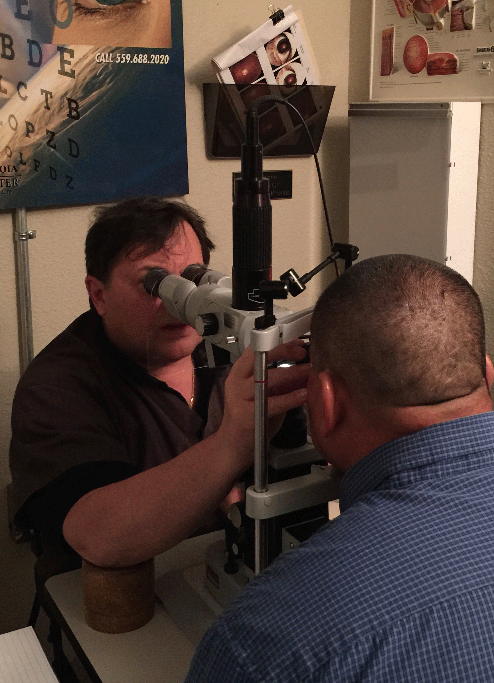
How is Diabetic Retinopathy Diagnosed?
If you are diagnosed with diabetes, it is very important for you to get annual eye exams from an ophthalmologist so diabetic retinopathy can be detected early. When you come for a visit we will ask you questions about your medical history. Because some of the signs of diabetic retinopathy cannot be seen during the basic eye exam, we will administer a few drops to dilate your pupils and Dr. Sunalp will directly examine your retina with lenses and equipment.
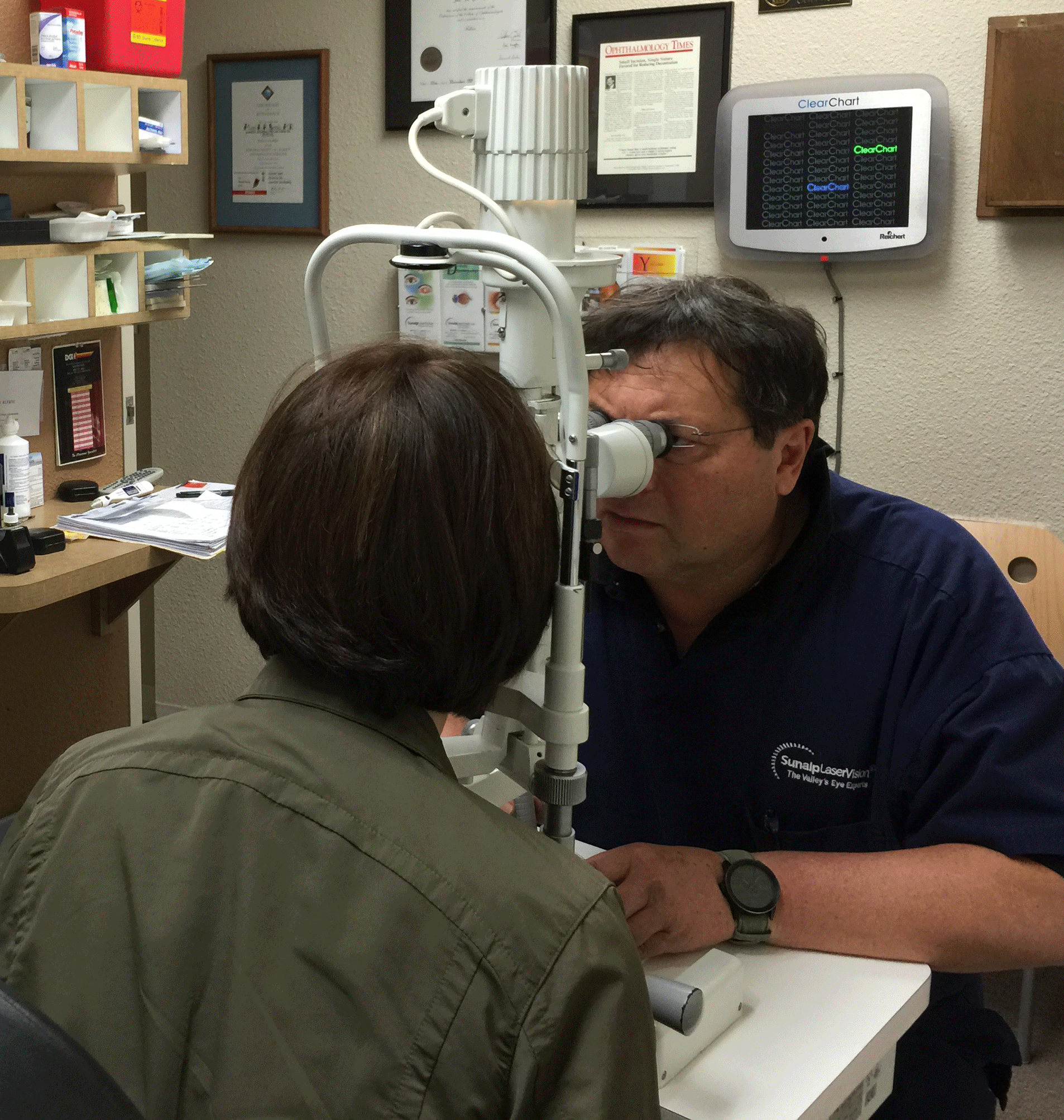
Dr. Sunalp performing a Slit-Lamp Exam
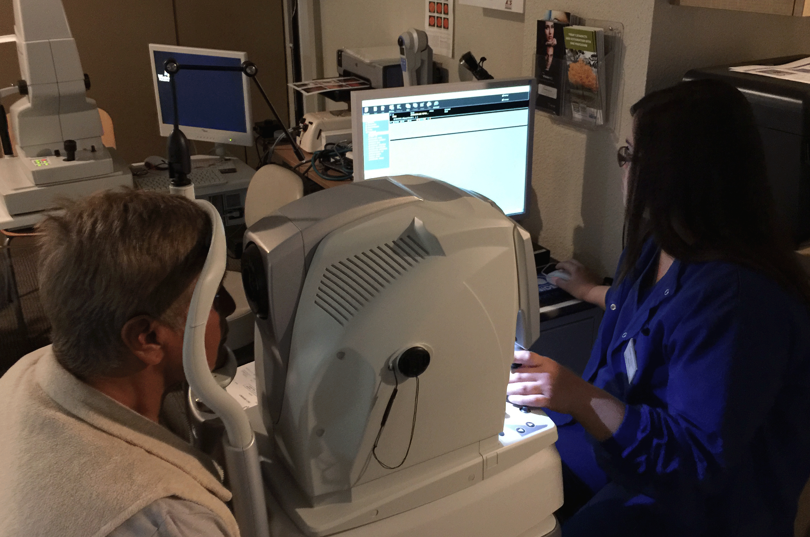
Optical coherence tomography (OCT) can be used to visualize details in the retina and is instrumental in diagnosing and monitoring the progression of Diabetic Retinopathy. At Sunalp Laser Vision we use the newest model of OCT, NIDEK-RS 300.
Normal Vision
Simulated Diabetic Retinopathy Vision
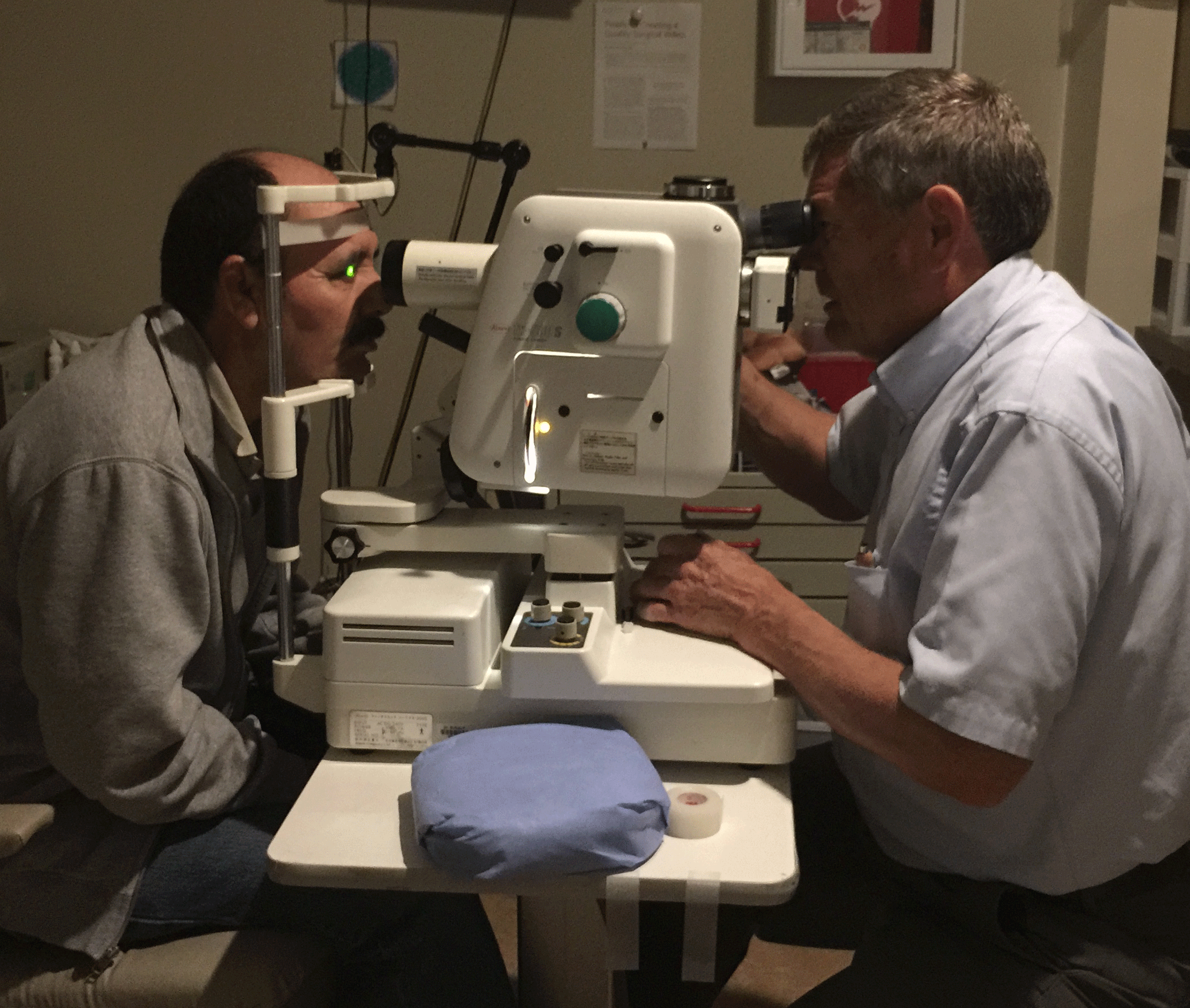
Fluorescein Angiography (FA) may be performed to further evaluate for any leaking blood vessels.
How is Diabetic Retinopathy Treated?
Treatment of diabetic retinopathy requires both a medical doctor and an ophthalmologist.
Your medical doctor will help you to maintain a better control of your blood sugar and also treat other complications associated with diabetic retinopathy (preserve kidney, heart and nerve function).
Your ophthalmologist can prevent further eye blood vessel changes and preserve your vision by using a laser treatment (photocoagulation).
Photocoagulation (laser treatment for diabetic retinopathy) is done by making very tiny, painless, retinal "burns" that seal off leaking blood vessels and reduce swelling.
Anti-VEGF therapy
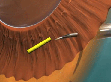
At Sunalp Laser Vision we use a highly successful treatment protocol involving use of a drug that impairs the production of new blood vessels. This class of drugs is called vascular endothelial growth factor (VEGF) inhibitors or anti-VEGF. In addition we use an innovative method of introducing this drug into the eye. This method was developed by Dr. Sunalp to specifically avoid many of the complications associated with the introduction of the drug into the eye. This treatment technique, which minimizes side effects while maximizing tretment benfits, is one of the many ways Sunalp Laser Vision has demonstrated commitment to state-of-the-art services.

Standard Approach

Dr. Sunalp's Perilimbic Approach
News on Diabetic Retinopathy Treatment
ILUVIEN Eye Implant for Diabetic Macular Edema (DME)
-
Approved by FDA for treatment of DME
-
Length of a grain of rice (3.5 mm x 0.37 mm)
-
Is injected intravitreally, using a syringe-like device
-
Once deployed, the device slowly releases fluocinolone acetonide (FAc), a corticosteroid, for the next three years to control inflammation within the eye and help slow down the effects of DME
-
Advantages of ILUVIEN over existing therapies is that a single injection provides sustained therapy for three years.
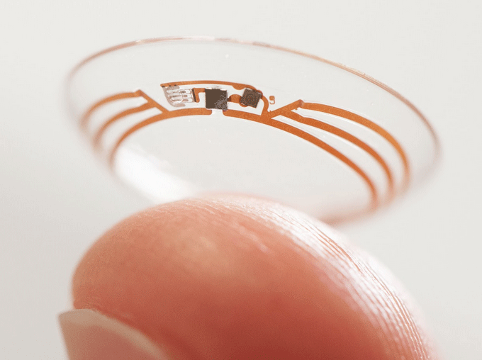
Google Contact Lens Glucose Monitor
Monitor diabetic's blood sugar levels by testing their tears
-
The contact lens uses processing chips and a glucose sensor
-
Next to the sensors lies an antenna thinner than a human hair
-
The sensors detect glucose levels in the wearer's tears, taking readings once per second, and the antenna transmits findings to an external device